What Causes Fibromyalgia?

The cause of fibromyalgia still remains unknown. However, several studies are in progress aiming to find out its cause. Significant progress has been made over the last decades regarding the pathogenesis of fibromyalgia. It seems that fibromyalgia constitutes a syndrome* sharing one broad symptomatology however not having one single cause.


*syndrome – is wider and more general term. It is set of signs and symptoms building clinical picture having various underlying causes. Disease in contrary has one specific cause usually.

symptomatology – (1) medical science dealing with disease symptoms. (2) set of symptoms related to specific medical condition.

Hormone Rhythm Abnormality

Sleep disorders are one of the most common among various symptoms of fibromyalgia. Fibromyalgia patients report early morning awakenings, awakening feeling tired or unrefreshed, insomnia, as well as mood and cognitive disturbances. Polysomnographic findings during sleep in these patients include an alpha frequency rhythm and presence of abnormal delta waves in non-REM sleep stage, so called alpha-delta sleep.

According to Bennett this could reflect in diminished production of growth hormone (GH) and insulin-like growth factor (IGF-1) that may cause improper muscle function and tiredness.

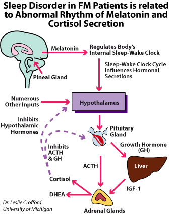
The effect of that (abnormal GH production) could be predisposition to micro-injuries in muscle fibres and inefficient repair mechanisms and subsequently weaker muscle function. The other authors suggest that the muscle weakness may result from poor energetic muscle reserve due to some amino acids like valine, leucinne, isoleucine, phenylalanine taking part in protein synthesis.

Changes in circadian rhythm are observed in vast majority of patients with fibromyalgia. Pain, stiffness and mood disturbances are common symptoms in fibromyalgia. All of these conditions have the features of disrupted sleep patterns and dysregulated biologic circadian rhythms, such as stress hormone secretion. Three hormones: cortisol, melatonin and DHEA play a key role in circadian rhythm. In the physiological condition cortisol has a very distinct circadian rhythm with its lowest level late night (2 – 4 hours after falling asleep) and highest in the late morning (3 – 4 hours after waking up).

The steroid hormones, cortisol and dehydroepiandrosterone (DHEA) are the two main peripheral secretory products of the hypothalamic-pituitary-adrenal stress-neuroendocrine axis. DHEA in many respects parallels cortisol secretory activity there was some dissociation; however mean levels were positively but not tightly correlated. The secretory pattern of DHEA seems very stable.

The melatonin is a hormone produced by the pineal gland and regulates sleep and wakefulness. Melatonin in contrary is secreted at night with a robust circadian rhythm and maximum plasma levels that occur around 3 to 4 AM. The daily rise of melatonin secretion correlates with a subsequent increase in sleep propensity about 2 hours before the person’s regular bedtime.

There is a hypothesis that the abnormal rhythm of hormone levels (cortisol, melatonin, and DHEA) is responsible for the changes of circadian rhythm of fibromyalgia patients. However, the total value of the daily secretion of these hormones remains in normal range.

Sleep disturbances have been more widely presented in bookmark → sleep disorders.

Serotonin Hypothesis

In view of the evidence regarding alterations of the serotoninergic system in the pathogenesis of fibromyalgia genetic research was initially directed towards genes involved in modulation of that system. Offenbaecher et al. compared the genotype of the serotonin transporter gene (5-HTT) promoter region in fibromyalgia patients with healthy controls. An increased frequency of the S/S genotype of the 5-HTT gene was demonstrated among patients versus controls.

These results were subsequently confirmed by a study analyzing Palestinian Arabs and Israeli Jews. On the other hand, another study directed at the T102C polymorphism of the 5-HT2A-receptor gene, another serotonin receptor candidate gene, failed to demonstrate a difference in the frequency of the polymorphism among fibromyalgia patients and controls.

Similarly, a study focusing on the serotonin receptor subunit genes, HTR3A and HTR3B found no significant difference in these genes among fibromyalgia patients.

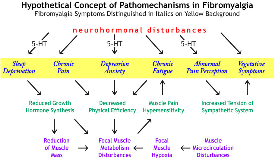
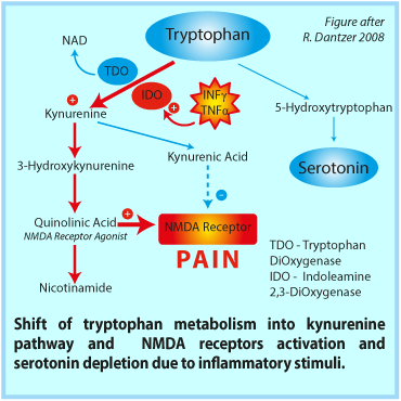
Possible links between neurochemical abnormalities and serotonin (5-HT) deficit and different FMS symptoms are presented in the figure beside. Click the link in order to magnify it → figure

Presence of 5-HT antibodies in serum and high density of serotonin receptors in nerve synapse are specified among various causes of 5-HT deficiency in fibromyalgia. Recently, a defined autoantibody pattern consisting of antibodies to serotonin (5-hydroxytryptamine, 5-HT), gangliosides and phospholipids was found in about 70% of the patients with fibromyalgia syndrome.

Kein and Berg investigated whether patients with chronic fatigue syndrome (CFS) express similar humoral immunoreactivity. Sera from 42 CFS patients were analysed by ELISA for these antibodies, and the results were compared with those previously observed in 100 FMS patients. 73% of the FMS and 62% of the CFS patients had antibodies to serotonin, and 71% or 43% to gangliosides, respectively. Antibodies to phospholipids could be detected in 54% of the FMS and 38% of the CFS patients. 49% of FMS and 17% of the CFS patients had all three antibodies in parallel, 70% and 55%, respectively had at least two of these antibody types. 21% of FMS and 29% of CFS patients were completely negative for these antibodies. Antibodies to 5-HT were closely related with FMS/CFS while antibodies to gangliosides and phospholipids could also be detected in other disorders. The observation that family members of CFS and FMS patients also had these antibodies represents an argument in favour of a genetic predisposition. These data support the concept that FMS and CFS may belong to the same clinical entity and may manifest themselves as 'psycho-neuro-endocrinological autoimmune diseases'.

Ref.:Klein R, Berg PA: High incidence of antibodies to 5-hydroxytryptamine, gangliosides and phospholipids in patients with chronic fatigue and fibromyalgia syndrome and their relatives: Evidence for a clinical entity of both disorders, Eur.J.Med.Res. 1(11):1995,21-26.


Impaired Pain Sensitsitization

Fibromyalgia is a disorder of pain processing. Evidence suggests that both the ascending and descending pain pathways operate abnormally, resulting in central amplification of pain signals, analogous to the “volume control setting” being turned up too high. Patients with FM also exhibit changes in the levels of neurotransmitters that cause augmented central nervous system pain processing; levels of several neurotransmitters that facilitate pain transmission are elevated in the cerebrospinal fluid and brain, and levels of several neurotransmitters known to inhibit pain transmission are decreased. Pharmacological agents that act centrally in ascending and/or descending pain processing pathways, such as medications with approved indications for FM, are effective in many patients with FM as well as other conditions involving central pain amplification. Research is ongoing to determine the role of analogous central nervous system factors in the other cardinal symptoms of FM, such as fatigue, nonrestorative sleep, and cognitive dysfunction.
Daniel J. Clauw, MD, Lesley M. Arnold, MD, and Bill H. McCarberg, MD: The Science of Fibromyalgia, Mayo Clin Proc. 2011 Sep; 86(9): 907–911.

The advent of imaging techniques capable of providing real time information regarding the way specific areas within the central nervous system react to painful (as well as non-painful stimulation) has provided valuable insight into the biological meaning of central sensitization in fibromyalgia. Thus, using functional MRI (fMRI), Gracely et al. were able to demonstrate that conditions which resulted in comparable subjective sensation of pain, resulted in activation patterns that were similar in fibromyalgia patients and controls; on the other hand, similar levels of pressure (which invoke higher levels of pain among fibromyalgia patients relative to controls) resulted in activation of different areas and caused greater effects in patients. Similarly Cook et al. showed that in response to painful stimuli, fibromyalgia patients had greater activity in the contralateral insular cortex than healthy controls, as demonstrated by fMRI.Thus, functional brain imaging such as fMRI has supplied fibromyalgia researches with a long-awaited tool for objective evaluation of pain. This tool enables further evaluation of various factors capable of influencing the functioning of the nervous system in fibromyalgia, ranging from pharmacological intervention to the presence of a significant other during testing.

It has not been found coherent evidence of the tissue pathology in fibromyalgia. Because of that the contemporary research focuses on exploration of the central (CNS) mechanisms of pain.

Click the figure to magnify

Central sensitization is an emerging biopsychosocial concept currently considered to characterize a wide spectrum of interrelated ‘functional’ disorders, which may subsequently be better defined as central sensitivity syndromes. Central sensitization constitutes a condition of general over reactivity of the central nervous system to a wide spectrum of stimulation. Clinical correlates which are particularly significant to the fibromyalgia syndrome include the phenomenon of allodynia, in which a normally un-painful stimulus is perceived of as painful and hyperalgesia, in which a normally painful stimulus is perceived of as more painful than expected. Additionally, central sensitization invokes prolonged electrophysiological discharge in response to stimulation as well as exaggerated response to various forms of stimulation such as noise, smell and chemical exposure. Various areas in the central nervous system are responsible for inhibiting ascending pain transmission within the spinal cord (e.g. the locus coeruleus*, cortico-reticular** system, brain stem and hypothalamus) through the activity of inhibitory neurotransmitters which include serotonin, norepinephrine, enkephalins, gamma-amino-butyric acid (GABA), and adenosine. A decrease in this pain inhibitory loop is an important component of the central sensitization syndrome. Separate areas of the central nervous system including the limbic system and the medial thalamic nuclei, are involved in the affective response of the central nervous system to pain.

Reference: Staud R, Smitherman ML. Peripheral and central sensitization in fibromyalgia: pathogenetic role. Curr Pain Headache Rep. 2002 Aug;6(4):259-66.


† Sensitization – an amplified response to a stimulus resulting from repeated exposure to it. Peripheral and central abnormalities of nociception have been described in fibromyalgia. Important nociceptor systems in the skin and muscles seem to undergo profound changes in patients with fibromyalgia through unknown mechanisms. They include sensitization of vanilloid receptor, acid-sensing ion channel receptors, and purino-receptors. Tissue mediators of inflammation and nerve growth factors can excite these receptors and cause extensive changes in pain sensitivity, but patients with fibromyalgia lack consistent evidence for inflammatory soft tissue abnormalities. Therefore, recent investigations have focused on central nervous system mechanisms of pain in fibromyalgia.

*The locus coeruleus is a nucleus in the pons of the brainstem involved with physiological responses to stress and panic. It is a part of the reticular activating system. The locus coeruleus is the principal site for brain synthesis of norepinephrine (noradrenaline). The locus coeruleus is responsible for mediating many of the sympathetic effects during stress. The locus coeruleus is activated by stress, and will respond by increasing norepinephrine secretion, which in turn will alter cognitive function (through the prefrontal cortex), increase motivation (through nucleus accumbens), activate the hypothalamic-pituitary-adrenal axis, and increase the sympathetic discharge/inhibit parasympathetic tone (through the brainstem). Specific to the activation of the hypothalamo-pituitary adrenal axis, norepinephrine will stimulate the secretion of corticotropin-releasing factor from the hypothalamus, that induces adrenocorticotropic hormone release from the anterior pituitary and subsequent cortisol synthesis in the adrenal glands. Norepinephrine released from locus coeruleus will feedback to inhibit its production, and corticotropin-releasing hormone will feedback to inhibit its production, while positively feeding to the locus coeruleus to increase norepinephrine production.

**The reticular formation is a set of interconnected nuclei that are located throughout the brainstem. It includes neurons located in diverse parts of the brain. The neurons of the reticular formation make up a complex set of networks in the core of the brainstem that stretches from the upper part of the midbrain to the lower part of the medulla oblongata. The reticular formation includes ascending pathways to the cortex in the ascending reticular activating system (ARAS) and descending pathways to the spinal cord via the reticulospinal tracts of the descending reticular formation.

Injury, Inflammation

Several papers have documented link between injury and fibromialgia. Buskila et al. have documented increased rates of fibromyalgia following cervical spine injury. A controlled study of 161 cases of traumatic injury. Arthritis Rheum. 1997 Mar;40(3):446-52.

B. Larsson et al. have reviewed Medline database and have found similar observations: A systematic review of risk factors associated with transitioning from regional musculoskeletal pain to chronic widespread pain. Eur J Pain. 2012 Sep;16(8):1084-93.

It has been shown that inflammation and/or injury may affect serotonin levels through activation of the kynurenine pathway. Pro-inflammatory cytokines activate the enzyme indoleamine 2,3-dioxygenase (IDO). IDO shuttles tryptophan, the precursor to 5-hydroxytryptophan (5-HTP), down the kynurenine pathway rather than down the pathway that would result in the synthesis of serotonin (see the figure). Activation of the kynurenine pathway can result in depletion of tryptophan and consequently, depletion of serotonin. Furthermore, 5-HTP and serotonin itself can be substrates for IDO leading to further serotonin depletion.

In addition to decreasing serotonin levels in the body, the kynurenine pathway also results in the synthesis of multiple metabolites that can have neurotoxic effects. These metabolites include 3-hydroxy-kynurenine (3-OH-KYN) as well as quinolinic acid. 3-OH-KYN exerts its damaging effects by increasing reactive oxygen species* (ROS) in the brain. This increase in ROS leads to oxidative stress and possibly neuronal apoptosis. Overproduction of ROS is linked to an increase in Monoamine oxidase (MAO) activity, which could deplete monoamine levels in the brain leading to a depressed mood. Quinolinic acid is a potent N-methyl-D-aspartate (NMDA) receptor agonist. Overstimulation of these receptors leads to an increased calcium influx into target neurons resulting in neuronal damage. Increased influx of calcium into cells may also lead to the generation of ROS. Both metabolites have been found to be elevated in certain neurodegenerative disorders such as Huntington’s disease and Parkinson’s disease


*Reactive Oxygen Species - ROS - are chemically reactive chemical species (other words chemical substances) containing oxygen. Examples include peroxides, superoxide, hydroxyl radical, and singlet oxygen.


Contact

If you would like to contact us

fibromyalgia.zone

Mailing address:
ul. Farbiarska 22
02-862 Warsaw

Phone:    +48 22 487 14 44

Mail:   mail@fibromyalgia.zone

Contact Form

If you have not found the answer you are looking for, please fill in this contact form and send it to our expert!

By ticking this checkbox I hereby agree for processing of my personal data contained in this contact form to receive an answer to my inquiry by electronic mail by the Administrator i.e. Phytomedica Poland Co. Ltd. 31/35 Ogrodowa Street, 00-893 Warsaw, Poland.

Enter the code shown in the picture
This is a captcha-picture. It is used to prevent mass-access by robots. (see: www.captcha.net)

Before sending the form, make sure the email address you typed is correct!